Flint Office
1303 S. Linden Rd., Suite D
Flint, MI 48532

As you grow older, you will start to notice more problems with your feet due to wear and tear. This may also happen because the skin will start to become thin and lose elasticity. Some signs of aging feet are regular aches and pains, bunion development, and clawed toes.
Fortunately, there are ways you can improve comfort, relieve pain, and maintain mobility in your feet. One of the best ways to deal with aging feet is to exercise. If you keep active, your muscles will become toned which will then strengthen the arches in the foot and stimulate blood circulation.
It is important that you practice proper foot care to protect your aging feet. You should wash your feet in warm water on an everyday basis. Afterward, the feet need to be dried well and it is important to dry between the toes. Your toenails should be trimmed and kept under control; nails that are poorly cut may become ingrown. At the end of each day, performing an inspection of your feet will allow you to detect any ailments in their early stages.
As you grow older, it becomes more important that you wear comfortable shoes. Your shoes should be secure, and they should provide decent arch support. If you are looking to buy a new pair of shoes, it is best to look for a pair that are made from a breathable material. It is also helpful to have shoes that have a bit of extra room at the top of the shoe, especially if you suffer from swollen feet.
The most common foot problems that elderly people will encounter are bunions, calluses, corns, hammertoes, heel pain, and foot problems related to diabetes. Some other issues include arch pain, tarsal tunnel syndrome, Achilles tendonitis, and Morton’s neuroma
An annual foot examination is a great way for you to ensure that you do not have any serious health problems with your feet. You should talk to a podiatrist about the available treatment options for whichever foot issue you are dealing with.

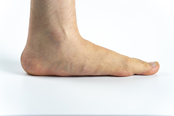 When the feet are pressed flat while standing, this can often be a result of the condition known as flat feet or fallen arches. Usually, flat feet are not serious and do not need treatment. They should not interfere with your daily physical activities either. However, in more serious cases, patients with flat feet should consult with a podiatrist. Common reasons a podiatrist should be consulted include flat feet that are painful, stiff, or weak. Patients who have flat feet and often injure their feet or ankles, have a balance problem, or have only one foot that is affected, should consult with a podiatrist as well. For certain cases of flat feet, surgery may be necessary, but in most cases, a podiatrist will be able to suggest proper footwear and exercises in order to treat this condition.
Flatfoot is a condition many people suffer from. If you have flat feet, contact one of our podiatrists from Community Podiatry Group. Our doctors will treat your foot and ankle needs.
What Are Flat Feet?
Flatfoot is a condition in which the arch of the foot is depressed and the sole of the foot is almost completely in contact with the ground. About 20-30% of the population generally has flat feet because their arches never formed during growth.
Conditions & Problems:
Having flat feet makes it difficult to run or walk because of the stress placed on the ankles.
Alignment – The general alignment of your legs can be disrupted, because the ankles move inward which can cause major discomfort.
Knees – If you have complications with your knees, flat feet can be a contributor to arthritis in that area.
Symptoms
Treatment
If you are experiencing pain and stress on the foot you may weaken the posterior tibial tendon, which runs around the inside of the ankle.
If you have any questions please feel free to contact our office located in Flint, MI . We offer the newest diagnostic and treatment technologies for all your foot and ankle needs.
Flatfoot is a foot condition in which the arch of the foot has either partially or totally dropped or has never developed. While it is common in babies and small children, it can become a problem for them in adulthood if the arch never forms. For adults, the development of flat feet can be brought upon by injury, as a result of pregnancy due to increased elasticity, or obesity. Those who have health concerns such as rheumatoid arthritis or diabetes may also be at greater risk for developing the condition.
If you suspect that you have flat feet, it is best to consult your podiatrist. Your foot doctor will examine the suspected foot and observe how it looks while you sit and stand. He or she may take an X-ray to determine how serious the condition is. Some common signs of flatfoot include toe drift, in which the toes and front part of the foot point outward, a short Achilles tendon, and a heel that tilts outwardly while the ankle tilts inward.
Once flatfoot has been diagnosed, your podiatrist may suggest one of several treatment options. Flat feet can be rigid, in which the feet appear to have no arch even when the person is not standing; or flexible, in which the person appears to have an arch while not standing, but once standing the arch disappears. Those with flexible flatfoot may be told to reduce any activities that cause pain and to avoid extended periods of walking or standing. Another suggestion may be weight loss, as excessive weight may be placing pressure on the arches
In few cases, if the condition is severe and all other methods have been exhausted surgery may be required. This is normally avoided, however, due to a lengthy recovery time and high cost.


Plantar warts are small growths that develop on parts of the feet that bear weight. They're typically found on the bottom of the foot. Don't live with plantar warts, and call us today!

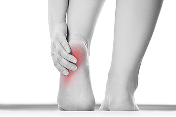
As the foundation of the body, the foot and ankle are complex, amazing structures made up of many ligaments, nerves, tendons, joints, and bones—including the largest bone in the foot, the heel. The heel helps form the arch and manages stress during walking and running. All of this use and stress can lead to painful conditions of the heel and its surrounding tendons and ligaments. Plantar fasciitis, the most common form of heel pain, is an inflammation of the connective tissue that spans the bottom of the foot (plantar fascia). Another common form of heel pain occurs with Achilles tendonitis, which causes inflammation and pain in the (Achilles) tendon that attaches the heel with the calf muscles. A loss of cushioning in the plantar fat pad can cause the heel to become bruised, and the heel bone can also fracture, causing intense pain. Retrocalcaneal Bursitis is an inflammation of the fluid-filled bursa sac that cushions the back of the heel bone. Any type of heel pain should be diagnosed by a podiatrist. who can assess the situation and create an appropriate treatment plan based on their findings.
Many people suffer from bouts of heel pain. For more information, contact one of our podiatrists of Community Podiatry Group. Our doctors can provide the care you need to keep you pain-free and on your feet.
Causes of Heel Pain
Heel pain is often associated with plantar fasciitis. The plantar fascia is a band of tissues that extends along the bottom of the foot. A rip or tear in this ligament can cause inflammation of the tissue.
Achilles tendonitis is another cause of heel pain. Inflammation of the Achilles tendon will cause pain from fractures and muscle tearing. Lack of flexibility is also another symptom.
Heel spurs are another cause of pain. When the tissues of the plantar fascia undergo a great deal of stress, it can lead to ligament separation from the heel bone, causing heel spurs.
Why Might Heel Pain Occur?
Treatments
Heel pain should be treated as soon as possible for immediate results. Keeping your feet in a stress-free environment will help. If you suffer from Achilles tendonitis or plantar fasciitis, applying ice will reduce the swelling. Stretching before an exercise like running will help the muscles. Using all these tips will help make heel pain a condition of the past.
If you have any questions please contact our office located in Flint, MI . We offer the newest diagnostic and treatment technologies for all your foot and ankle needs.
Heel pain can be difficult to deal with, especially if you do not know what the underlying cause is. If you ignore your heel pain, the pain can magnify and potentially develop into a chronic condition. Depending on the location of your heel pain, you have developed a specific condition.
One condition is plantar fasciitis. Plantar fasciitis is caused by the inflammation of the plantar fascia, or the band of tissue that connects the heel bone to the base of the toes. The pain from this condition is initially mild but can intensify as more steps are taken when you wake up in the morning. To treat this condition, medication will likely be necessary. Plantar fasciitis is often associated with heel spurs; both require rest and special stretching exercises.
There are various options your podiatrist may suggest for heel pain. Treatment options for heel pain typically include non-steroidal anti-inflammatory drugs (NSAIDS), which may reduce swelling and pain. Other options are physical therapy, athletic taping, and orthotics. In severe cases of heel pain, surgery may be required.
Preventing heel pain is possible. If you are looking to prevent heel pain from developing in the future, be sure to wear shoes that fit you properly and do not have worn down heels or soles. Be sure to warm up properly before participating in strenuous activities or sports that place a lot of a stress on the heels. If you are experiencing any form of heel pain, speak with your podiatrist to determine the underlying cause and receive the treatment you need.
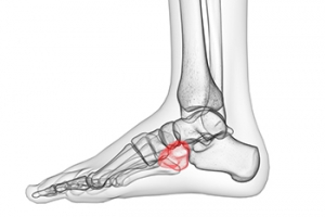

The cuboid bone is a tiny, cube-shaped (cuboidal) bone, which is one of the seven tarsal bones of the foot. It is located on the outside of the foot, roughly halfway between the pinky toe and the heel. The cuboid bone moves slightly during normal foot motion, however, forceful movements or certain positions that are maintained for prolonged periods of time can cause the cuboid bone to shift excessively and therefore it becomes dislocated. This is known as cuboid syndrome. Symptoms may include pain and possibly swelling on the lateral (outside) part of the foot which increases when walking or standing. It may even be difficult or impossible to walk. Podiatrists may use a “whip” procedure to quickly and firmly apply force to the cuboid bone to get it back into alignment as the patient relaxes in a prone position. If you believe you may be suffering from cuboid syndrome, contact a podiatrist.
Cuboid syndrome, also known as cuboid subluxation, occurs when the joints and ligaments near the cuboid bone in the foot become torn. If you have cuboid syndrome, consult with one of our podiatrists from Community Podiatry Group. Our doctors will assess your condition and provide you with quality foot and ankle treatment.
Cuboid syndrome is a common cause of lateral foot pain, which is pain on the outside of the foot. The condition may happen suddenly due to an ankle sprain, or it may develop slowly overtime from repetitive tension through the bone and surrounding structures.
Causes
The most common causes of cuboid syndrome include:
Symptoms
A common symptom of cuboid syndrome is pain along the outside of the foot which can be felt in the ankle and toes. This pain may create walking difficulties and may cause those with the condition to walk with a limp.
Diagnosis
Diagnosis of cuboid syndrome is often difficult, and it is often misdiagnosed. X-rays, MRIs and CT scans often fail to properly show the cuboid subluxation. Although there isn’t a specific test used to diagnose cuboid syndrome, your podiatrist will usually check if pain is felt while pressing firmly on the cuboid bone of your foot.
Treatment
Just as the range of causes varies widely, so do treatments. Some more common treatments are ice therapy, rest, exercise, taping, and orthotics.
If you have any questions, please feel free to contact our office located in Flint, MI . We offer the newest diagnostic and treatment technologies for all your foot care needs.
Cuboid syndrome mostly affects athletes, although it can affect non-athletes too. It is also known as cuboid subluxation or cuboid fault syndrome. This condition occurs when joints and ligaments near the cuboid bone of the foot are damaged, or when the cuboid bone itself is dislodged from its natural position. It is usually marked by pain on the outer side of the foot, which may be persistent or may come and go. Cuboid syndrome can be difficult to diagnose unless it becomes severe and more noticeable. Your doctor will likely ask questions about when the pain began and how long it has been present, and will put pressure on the cuboid bone to determine if that area is the origin of the pain.
Causes of Cuboid Syndrome
Disagreements Amongst Podiatrists Regarding Cuboid Syndrome
It is very important that when you experience any kind of pain on the side of your foot, you should seek medical care right away. If a subluxed cuboid is caught early, your feet may respond well to the treatment, and you can get back into sports or other activities again as soon as the pain subsides.

Vascular diseases affect the circulatory system and can involve blood disorders or abnormalities in the arteries, veins, or lymph vessels. Peripheral Artery Disease (PAD) is a type of vascular disease which causes a narrowing or blockage in the arteries that prevents their capacity to carry oxygen-rich blood away from the heart to the legs and feet. PAD can cause leg pain when walking (claudication), numbness, tingling, coldness, or an inability for wounds to heal in the legs or feet. Left untreated, PAD may also be a precursor to life-threatening issues such as a heart attack or stroke. If you are experiencing any of these symptoms in your legs or feet, schedule an appointment with a podiatrist who can help diagnose and treat PAD. They will perform a physical examination and may even suggest that tests be performed to help assess the presence and severity of vascular disease, such as a Computed Tomography Angiography (CTA) and Magnetic Resonance Angiography (MRA). CTA uses a type of X-ray scan, while MRA uses radio wave technology. Both tests produce 3D imaging and typically involve inserting a contrasting material (dye) in the blood vessels which makes them easier to visualize.
Vascular testing plays an important part in diagnosing disease like peripheral artery disease. If you have symptoms of peripheral artery disease, or diabetes, consult with one of our podiatrists from Community Podiatry Group. Our doctors will assess your condition and provide you with quality foot and ankle treatment.
What Is Vascular Testing?
Vascular testing checks for how well blood circulation is in the veins and arteries. This is most often done to determine and treat a patient for peripheral artery disease (PAD), stroke, and aneurysms. Podiatrists utilize vascular testing when a patient has symptoms of PAD or if they believe they might. If a patient has diabetes, a podiatrist may determine a vascular test to be prudent to check for poor blood circulation.
How Is it Conducted?
Most forms of vascular testing are non-invasive. Podiatrists will first conduct a visual inspection for any wounds, discoloration, and any abnormal signs prior to a vascular test.
The most common tests include:
These tests are safe, painless, and easy to do. Once finished, the podiatrist can then provide a diagnosis and the best course for treatment.
If you have any questions, please feel free to contact our office located in Flint, MI . We offer the newest diagnostic and treatment technologies for all your foot care needs.
In foot care, vascular testing may be required in the diagnosing and treatment of certain podiatric conditions. Vascular testing is particularly relevant for patients with high-risk diabetes, poor circulation, peripheral artery disease (PAD), and chronic venous insufficiency (CVI). Procedures typically involve the examination of blood vessels throughout the body for blockages or buildup.
Vascular testing is very important for the diagnosis of various conditions, including peripheral artery disease and chronic venous insufficiency, as these conditions can greatly affect one’s quality of life and cause pain in the lower limbs. Circulatory problems in the feet and ankles can reflect issues throughout the body, making testing of the blood vessels pertinent.
Testing methods vary between practitioners and can be specific to certain foot and ankle problems. Modern technology has brought about the ability to perform vascular testing using non-invasive methods, such as the cuff-based PADnet testing device. This device records the Ankle-Brachial Index (ABI)/Toe-Brachial Index (TBI) values and Pulse Volume Recording (PVR) waveforms. Contact your podiatrist to determine what vascular testing is available for your needs.